
No products in the cart.
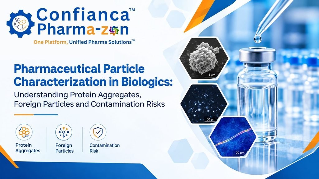
Pharmaceutical Particle Characterization in Biologics: Understanding Protein Aggregates, Foreign Particles and Contamination Risks
Introduction
Biopharmaceutical products have transformed modern healthcare through the development of monoclonal antibodies, recombinant proteins, vaccines and other advanced therapeutic products. However, their complex molecular structures and sensitive manufacturing processes create unique challenges for pharmaceutical quality teams.
One important challenge is the presence of particulate matter.
Unlike conventional small-molecule pharmaceutical products, biologics may contain particles originating from protein aggregation, product degradation, manufacturing equipment, packaging components, processing materials or external contamination.
Understanding the origin and characteristics of these particles is essential for maintaining product quality, patient safety and manufacturing consistency.
Pharmaceutical particle characterization provides a scientific approach to investigating particulate matter through physical, morphological, chemical and elemental analysis.
This article explains how pharmaceutical manufacturers can approach particle characterization in biologics, distinguish protein-related particles from foreign contaminants and develop scientifically supported investigation strategies.
1. Why Particle Characterization Matters in Biopharmaceutical Manufacturing
Biological medicines are particularly sensitive to environmental and processing conditions.
Factors such as temperature fluctuations, agitation, freezing and thawing, exposure to interfaces and formulation instability can influence particle formation.
Particle-related concerns may include:
- Protein aggregation during manufacturing or storage.
- Foreign particulate contamination from processing equipment.
- Glass particles originating from pharmaceutical containers.
- Elastomeric particles from closures and product-contact components.
- Fibres from garments, filters or environmental sources.
- Silicone-related particles associated with certain delivery systems.
Not every particle has the same origin or quality implication.
For example, protein aggregates and foreign inorganic particles require different analytical approaches. Therefore, identifying particle composition before assigning a root cause is important.
2. Understanding Different Types of Particles in Biologics
A pharmaceutical investigation should begin by classifying the observed particulate matter.
A. Protein aggregates
Protein aggregation can occur when proteins undergo structural changes or interact under unfavourable conditions.
Potential contributing factors include:
- Temperature variations.
- Mechanical stress.
- Air-liquid interfaces.
- Improper storage conditions.
- Formulation instability.
- Repeated freeze-thaw cycles.
Protein aggregates may be soluble or insoluble and can differ significantly in size and structure.
B. Inorganic particles
Inorganic particles may originate from glass, metals, mineral materials or other processing-related sources.
Elemental analysis can help identify characteristic elements and support comparisons with suspected materials.
C. Polymer and elastomer particles
These particles may originate from tubing, seals, stoppers, gaskets, bags and other product-contact components.
FTIR spectroscopy can be useful for identifying polymeric materials when the particle size, sample condition and analytical method permit.
D. Fibres and environmental contaminants
Fibres can originate from garments, wipes, packaging materials and the manufacturing environment.
Their morphology and chemical composition can help narrow potential sources.
3. Selecting the Right Analytical Technique
A reliable pharmaceutical particle characterization strategy should use analytical techniques according to the nature of the sample and the investigation objective.
Optical microscopy
Optical microscopy provides initial information about:
- Particle shape.
- Colour.
- Surface appearance.
- Approximate dimensions.
- Particle distribution.
It is often an appropriate starting point for documenting visible particles and selecting representative particles for further analysis.
Scanning Electron Microscopy (SEM)
SEM provides detailed morphological information at higher magnification.
It helps investigators examine surface structures, particle morphology and physical characteristics that may not be visible through conventional microscopy.
Energy Dispersive X-ray Spectroscopy (EDS)
SEM-EDS supports elemental characterization.
It can help identify elements such as iron, silicon, aluminium, calcium, chromium and other detectable elements within an analysed particle.
However, elemental composition alone does not always establish the exact material or source.
Fourier Transform Infrared Spectroscopy (FTIR)
FTIR provides molecular fingerprint information and is useful for investigating many organic materials, including polymers and fibres.
The suitability of FTIR depends on particle dimensions, material composition and sample preparation.
Complementary analytical techniques
Depending on the investigation, techniques such as Raman spectroscopy, micro-FTIR, light obscuration and flow imaging may provide additional information.
The selected method should answer a defined analytical question rather than simply generate more data.
4. A Practical Workflow for Investigating Particles in Biologics
A structured investigation improves consistency and scientific defensibility.
Step 1: Preserve the original sample
Document the product, batch, container, sampling location and observed condition. Avoid unnecessary handling that may introduce secondary contamination.
Step 2: Perform preliminary visual examination
Record particle colour, shape, approximate dimensions and distribution.
Step 3: Select representative particles
Where multiple particles are available, select representative particles based on morphology and other relevant characteristics.
Step 4: Conduct appropriate instrumental analysis
Choose SEM-EDS, FTIR or complementary methods according to the suspected material and analytical objective.
Step 5: Compare analytical findings
Compare results with potential manufacturing, packaging and environmental sources.
Step 6: Evaluate uncertainty
Consider sample preparation, substrate contribution, particle heterogeneity, detection limitations and instrument settings.
Step 7: Prepare a scientific investigation report
Present observations, analytical results, limitations and conclusions separately.
5. Why Sample Preparation Can Influence Results
Sample preparation is an important part of pharmaceutical particle characterization.
Improper handling can change particle morphology, introduce contamination or affect the reliability of analytical findings.
Important considerations include:
- Selecting a suitable collection membrane.
- Using appropriate tools for particle recovery.
- Avoiding contact with potential contaminating materials.
- Documenting the original sample condition.
- Evaluating substrate background during elemental analysis.
- Maintaining sample traceability.
For SEM-EDS analysis, the substrate should be evaluated carefully because its elemental contribution may influence the spectrum.
For FTIR analysis, substrate selection and background interference should be considered before interpreting the resulting spectrum.
6. Connecting Analytical Results with Manufacturing Sources
Identifying a material is only one part of a successful contamination investigation.
The next step is evaluating whether the material is consistent with a potential source.
For example:
| Analytical observation | Potential investigation area |
|---|---|
| Iron-rich particle | Stainless steel equipment or metallic components |
| Silicon-rich particle | Glass, silicone or mineral-related materials |
| Polymer-like spectrum | Tubing, seals, bags or packaging |
| Cellulose-like fibre | Wipes, garments or paper-based materials |
| Calcium-rich inorganic material | Raw materials, process residues or environmental sources |
These are investigative possibilities, not definitive source assignments.
A reliable conclusion should combine analytical findings with equipment history, maintenance records, manufacturing observations and material comparisons.
7. Documentation and Quality Risk Assessment
A pharmaceutical particle characterization report should contain sufficient information to support independent technical review.
Recommended report elements include:
- Sample identification and traceability.
- Description of the particle.
- Optical or SEM micrographs.
- Analytical methodology.
- Elemental composition or molecular identification results.
- Relevant spectra and supporting data.
- Analytical limitations.
- Potential source assessment.
- Investigation conclusion.
- Recommendations for further investigation, where necessary.
Risk assessment should consider product type, route of administration, particle characteristics, exposure and relevant regulatory expectations.
Analytical identification should not automatically be treated as proof of patient risk or proof of contamination origin.
8. How Confianca Pharmazon Supports Particle Characterization
Confianca Pharmazon supports pharmaceutical quality and contamination investigation requirements through particle characterization-related solutions and analytical services.
Our focus includes:
- Particle identification and characterization.
- SEM-EDS elemental analysis.
- Microscopy-based particle examination.
- Particle morphology assessment.
- Analytical reporting.
- Technical support for pharmaceutical contamination investigations.
Our objective is to help pharmaceutical professionals obtain meaningful analytical information to support quality investigations and scientifically justified decisions.
Frequently Asked Questions
1. What is pharmaceutical particle characterization?
It is the scientific examination of particulate matter to understand its physical, morphological, chemical and elemental properties.
2. Why is particle characterization important in biologics?
It helps distinguish protein-related particles from foreign contaminants and supports quality investigations.
3. Can SEM-EDS identify protein aggregates?
SEM-EDS provides elemental information and morphology, but it is generally not sufficient by itself to confirm protein identity. Complementary molecular techniques may be required.
4. Which technique is suitable for polymer particle identification?
FTIR or Raman spectroscopy may be suitable, depending on particle size, material characteristics and sample condition.
5. Does identifying a particle confirm its contamination source?
No. Identification provides evidence about material composition. Source attribution requires additional manufacturing and material-comparison evidence.
6. What information should a particle characterization report include?
It should include sample traceability, microscopy, analytical results, methodology, limitations, interpretation and conclusions.
Conclusion
Biopharmaceutical manufacturing requires a scientific and systematic approach to particulate matter investigation.
Understanding whether a particle originates from protein aggregation, manufacturing equipment, packaging materials or an external source is essential for developing meaningful quality investigations.
Pharmaceutical particle characterization combines microscopy, elemental analysis and molecular identification to provide a stronger technical foundation for contamination assessment.
Confianca Pharmazon aims to support pharmaceutical manufacturers and quality professionals with practical particle characterization solutions and technical expertise.
Need support with pharmaceutical particle identification or contamination investigation? Connect with Confianca Pharmazon.