
No products in the cart.
Particle Characterization in Pharmaceuticals: A Practical QA/QC Investigation Guide
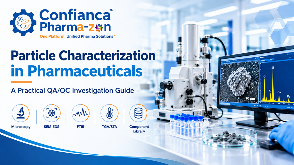
Particle characterization in pharmaceuticals helps quality assurance (QA), quality control (QC), and manufacturing teams investigate unexpected particles or fibers found in pharmaceutical products, process materials, or packaging systems. A useful investigation goes beyond naming a particle: it documents the sample, selects suitable analytical methods, evaluates the evidence, and considers plausible sources without overstating what the data can prove.
This guide focuses on how to plan and interpret a particle investigation. For service scope and sample discussions, see Confianca Pharmazon’s Particle Characterization Analysis Services.
What is particle characterization in pharmaceuticals?
Particle characterization is the systematic examination of an observed particle’s physical appearance and, where appropriate, its chemical or elemental properties. Depending on the sample and investigation question, it may help distinguish materials such as glass, metal, fibers, elastomeric fragments, polymers, or other process-related matter. The appropriate conclusion depends on the evidence collected and the limitations of each technique.
Characterization is not the same as proving the exact source. A result may be consistent with a component material, but source attribution normally requires additional comparison with relevant components, process knowledge, and investigation records.
Why particle investigations need a structured approach
- Product-quality assessment: establish what was observed and where it was found.
- Root-cause investigation: compare analytical findings with plausible materials and process sources.
- Evidence preservation: document sample handling and recovery so the investigation remains traceable.
- Risk-based decisions: provide QA and technical teams with clear results, uncertainty, and limitations.
- CAPA support: help inform follow-up checks, source verification, and corrective or preventive actions.
Step 1: Define the question before selecting a method
Begin by recording the product and sample type, the location and circumstances in which the particle was observed, the number of samples available, and any handling or storage conditions that may affect the evidence. Preserve photographs and relevant batch, equipment, packaging, and deviation information. Avoid unnecessary manipulation of the particle before the analytical plan is agreed.
Define the investigation question clearly: Is the priority to document morphology, assess elemental composition, investigate an organic material, compare a particle with a suspected component, or build a reference library of product-contact materials? Different questions may require different methods.
Step 2: Recover and document the particle carefully
Recovery should be selected for the sample and particle type. Filtration may be suitable in some liquid-sample investigations, while other samples may need a different preparation route. Record the method used, relevant filters or tools, and any preparation limitations. Include appropriate controls where the investigation design calls for them, particularly when handling or preparation could introduce background material.
Before advanced analysis, document visible characteristics such as shape, color, transparency, surface texture, and apparent structure. Microscopy images provide useful context for later analytical results and comparisons.
Step 3: Select analytical methods based on the material question
No single technique answers every particle-identification question. Method selection should reflect the particle’s size, amount, physical condition, suspected composition, and the information required.
- Stereo or optical microscopy: supports visual examination, morphology documentation, and selection of areas for further analysis.
- Fluorescence microscopy or imaging: may provide additional contrast or fluorescence-related information for suitable samples.
- Scanning electron microscopy with energy-dispersive X-ray spectroscopy (SEM-EDS): SEM documents surface morphology at higher magnification, while EDS provides elemental information from the analyzed region. Elemental results do not, by themselves, establish a unique compound or prove the source.
- FTIR spectroscopy: can help assess the molecular characteristics of suitable organic or polymeric materials when the sample and method are appropriate.
- Other complementary techniques: methods such as EDXRF or thermal analysis may be considered when the material question, sample quantity, and analytical objective justify their use.
These techniques are not interchangeable, and not every sample requires every method. A staged approach can preserve limited material and avoid unnecessary testing.
Step 4: Interpret the results as a combined evidence set
Interpretation should connect the analytical observations with the original question. For example, microscopy may describe a particle’s morphology, while SEM-EDS may show that the analyzed region contains particular elements. That combination can narrow possible material classes, but it should not be presented as definitive identification when the evidence is inconclusive.
Consider potential interferences, substrate or filter contributions, mixed-material particles, coatings, and the limits of the analyzed area. Report observations separately from interpretations. If the data support several plausible explanations, state them and explain what further comparison would help distinguish them.
Step 5: Compare findings with relevant component references
When source investigation is required, compare the particle’s characteristics with suitable reference materials from the product-contact or manufacturing environment. Reference work may involve identifying relevant components, collecting representative samples or coupons, and characterizing those materials with methods appropriate to their composition. A library is useful only when its entries are traceable and representative of the materials and components being considered.
A match in one characteristic should be treated cautiously. Stronger source hypotheses generally rely on multiple compatible observations, suitable reference samples, process context, and documented reasoning.
For a specific example of a focused investigation, read Glass Particle in an Injectable: Critical Steps for Source Investigation and Fiber Contamination in Injectable Products.
Step 6: Prepare a clear, traceable report
A useful analytical report should make the evidence understandable to both technical reviewers and quality decision-makers. Depending on the work performed, reporting may include:
- Sample identification and the stated investigation objective.
- Recovery or preparation approach and relevant limitations.
- Microscopy observations and representative images.
- SEM images, EDS spectra, and elemental results where performed.
- Other analytical results when the selected methods support them.
- Comparison with reference materials, including the basis for comparison.
- Interpretation, uncertainty, limitations, and recommended follow-up questions.
Reports should not imply that a method was performed if it was not, or that a source has been proven when the findings only indicate compatibility.
Common mistakes to avoid
- Choosing an analytical technique before defining the investigation question.
- Handling or preparing the sample without recording the steps.
- Interpreting elemental composition as a unique chemical identity.
- Attributing a particle to a component based on a superficial or incomplete match.
- Ignoring background contributions, sample limitations, or conflicting evidence.
- Writing conclusions more strongly than the data support.
Frequently asked questions
What is the purpose of particle characterization in pharmaceuticals?
It helps describe and assess unexpected particles through appropriate microscopy and analytical methods, supporting a documented investigation into material identity and possible sources.
Which technique is best for identifying an unknown pharmaceutical particle?
There is no universally best technique. Microscopy, SEM-EDS, FTIR, and other methods provide different types of information. Selection depends on the sample, suspected material, particle size, and the question being investigated.
Can SEM-EDS identify the exact source of a particle?
SEM-EDS can provide morphology and elemental information, which may narrow possible material classes. It does not automatically prove a unique material identity or exact source. Comparison with suitable references and process evidence may be needed.
Why are reference libraries useful?
Traceable reference libraries provide comparison data for relevant components and materials. Their value depends on representative sampling, suitable analytical methods, and careful interpretation.
What should a particle-characterization report contain?
It should document the sample, methods actually used, relevant images and analytical data, reference comparisons where available, interpretation, and limitations.
Discuss a particle-characterization investigation
Confianca Pharmazon supports pharmaceutical particle-characterization investigations using suitable recovery, microscopy, and analytical approaches, including SEM-EDS where appropriate. The available approach depends on the sample and the investigation objective.
To discuss sample suitability and the information required for an investigation, visit Particle Characterization Analysis Services or contact support@confiancapharmazon.com.