
No products in the cart.
SEM-EDS vs FTIR for Particle Characterization: Which Technique Is Best for Pharmaceutical Analysis?
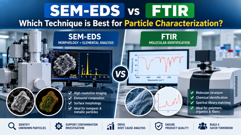
When an unknown particle is discovered in a pharmaceutical product, identifying it is only the beginning.
The more important question is often:
Where did the particle come from, and what does its identity tell us about the manufacturing process?
For pharmaceutical manufacturers, quality-control laboratories and investigation teams, two analytical techniques frequently considered for this purpose are Scanning Electron Microscopy with Energy Dispersive X-ray Spectroscopy (SEM-EDS) and Fourier Transform Infrared Spectroscopy (FTIR).
Both can provide valuable information, but they answer different analytical questions.
So, when comparing SEM-EDS vs FTIR for particle characterization, which one is better?
The answer is: neither technique is universally better. The right technique depends on the particle, the information required, and the investigation objective.
SEM-EDS is particularly valuable for high-resolution morphology and elemental composition. FTIR can provide molecular and chemical-structure information and is especially useful for many organic and polymeric materials. Published pharmaceutical investigations have demonstrated that combining complementary analytical techniques can be highly effective for identifying foreign matter.
This guide explains the differences between SEM-EDS and FTIR, when to use each, their advantages and limitations, and why a combined particle characterization analysis approach can be particularly useful for pharmaceutical contamination investigations.
Why Particle Characterization Matters in Pharmaceutical Manufacturing
Particles found in pharmaceutical products can originate from numerous sources.
For example, an unexpected particle could potentially be associated with:
- Manufacturing equipment
- Stainless-steel components
- Elastomeric components
- Polymer parts
- Tubing
- Filters
- Glass containers
- Packaging components
- Fibers
- Maintenance activities
- Environmental sources
- Process residues
Simply observing a particle under normal lighting may provide information about its color, shape and approximate appearance, but this may not be enough to determine its material composition or source.
This is where pharmaceutical particle characterization becomes valuable.
A structured investigation can move through several stages:
Detect → Recover → Observe → Characterize → Identify → Compare → Investigate Source
Analytical characterization can therefore transform an unknown particle from a visual observation into a source-investigation opportunity.
Research on pharmaceutical foreign matter has demonstrated the value of combining microscopy, SEM/EDS and FTIR because each technique contributes different information.
SEM-EDS vs FTIR: The Basic Difference
Before comparing the two technologies in detail, it helps to understand the fundamental difference.
What Does SEM-EDS Tell You?
SEM provides high-resolution images of the particle’s morphology and surface.
EDS provides information about the elements present in the analyzed area.
Together, SEM-EDS can answer questions such as:
- What does the particle look like at high magnification?
- Is it metallic or inorganic?
- Which elements are present?
- Does the particle contain iron, chromium, silicon, aluminum or other elements?
- Does its elemental profile resemble a manufacturing component?
What Does FTIR Tell You?
FTIR analyzes how a material interacts with infrared radiation and provides a spectrum related to its molecular structure and functional groups.
It can be particularly useful for:
- Polymers
- Elastomers
- Organic materials
- Certain residues
- Fibers
- Other materials with useful infrared signatures
FTIR can also be combined with spectral-library searching to help identify unknown materials. Research on pharmaceutical foreign-material investigations describes FTIR as particularly useful for obtaining chemical-structure information.
In simple terms:
SEM-EDS → morphology + elemental information
FTIR → molecular/chemical information
That distinction is the foundation of the SEM-EDS vs FTIR for particle characterization comparison.
How SEM-EDS Works in Particle Characterization
Scanning Electron Microscopy
SEM uses a focused electron beam to scan the sample surface and generate highly detailed images.
This allows analysts to examine particle characteristics that may not be visible using conventional microscopy.
SEM can provide information about:
- Particle morphology
- Surface structure
- Shape
- Texture
- Fracture patterns
- Particle size characteristics
- Surface features
This can be particularly useful when investigating small or irregular particles.
One pharmaceutical forensic-analysis publication notes that light microscopy and SEM provide morphological information, while EDS contributes elemental analysis.
Energy Dispersive X-ray Spectroscopy
EDS complements SEM by providing elemental information.
When the electron beam interacts with the sample, characteristic X-rays can be generated. The EDS detector analyzes these signals to determine which elements are present.
For example, an unknown particle may show an elemental profile consistent with:
- Iron-based material
- Aluminum-containing material
- Silicon-rich material
- Calcium-containing material
- Other inorganic compositions
This information can be compared with candidate source materials.
Example
Suppose a particle recovered from an injectable product appears dark and metallic.
Visual examination alone may not establish its identity.
SEM may reveal an irregular fragment with a particular surface morphology.
EDS may then demonstrate an elemental composition containing iron and chromium.
The investigation team can compare this result with relevant stainless-steel components used in the manufacturing process.
That does not automatically prove the source, but it can significantly narrow the investigation.
How FTIR Works in Particle Characterization
FTIR takes a different analytical approach.
Instead of primarily asking:
“Which elements are present?”
FTIR asks:
“What molecular or chemical characteristics does this material exhibit?”
Different chemical structures produce different infrared absorption patterns.
This makes FTIR particularly useful when investigating organic and polymeric materials.
Potential examples include:
- Rubber
- Silicone-based materials
- Plastic components
- Polymer fragments
- Fibers
- Organic residues
Published pharmaceutical investigations have shown that FTIR can help identify unknown foreign materials by comparing spectral signatures with library standards.
SEM-EDS vs FTIR: Key Comparison
| Factor | SEM-EDS | FTIR |
|---|---|---|
| Primary information | Morphology + elemental composition | Molecular/chemical information |
| Particularly useful for | Metals and inorganic particles | Polymers, organics and many fibers |
| High-resolution imaging | Excellent | Depends on FTIR configuration |
| Elemental identification | Yes | No |
| Molecular fingerprint | Limited | Strong |
| Surface morphology | Excellent | More limited |
| Spectral library matching | Not the primary approach | Commonly useful |
| Polymer identification | Can provide supporting information | Often highly useful |
| Metal identification | Highly useful | Generally less suitable |
| Unknown particle investigation | Highly useful | Highly useful |
| Best approach | Complementary with other methods | Complementary with other methods |
The comparison shows why choosing one technique solely because it is “more advanced” can be misleading.
The analytical question should determine the technique.
When Is SEM-EDS Better?
SEM-EDS can be especially useful when the investigation involves metallic or inorganic particles.
Consider these examples:
Example 1: Suspected stainless-steel particle
A particle is found during inspection of an injectable product.
The manufacturing process uses stainless-steel equipment.
SEM provides high-resolution morphology, while EDS can provide an elemental profile that may be compared with the equipment material.
Example 2: Suspected glass particle
A particle is suspected to originate from a glass vial.
SEM-EDS can provide morphological and elemental information that may help determine whether the particle is consistent with a glass-based material.
Recent research has demonstrated the complementary use of optical microscopy, SEM-EDS and FTIR to characterize thin glass particulates associated with parenteral drug products.
Example 3: Equipment wear investigation
If a facility suspects mechanical wear, elemental information can be particularly valuable when comparing the particle against metal components.
When Is FTIR Better?
FTIR can be particularly useful when the suspected particle is organic, polymeric or elastomeric.
Example 1: Rubber particle
A dark particle is recovered from a pharmaceutical product.
If the manufacturing process contains elastomeric seals or gaskets, FTIR may provide useful molecular information for comparison.
Example 2: Polymer fragment
A particle appears translucent or plastic-like.
FTIR can help determine whether the material has characteristics consistent with a specific polymer family.
Example 3: Fiber investigation
A visible fiber is detected during an investigation.
Microscopy can establish morphology, while FTIR may provide useful information about the material’s chemical characteristics.
This combination can help distinguish different types of fibers and identify potential sources.
Why Combining SEM-EDS and FTIR Can Be More Powerful
The real question is not always:
SEM-EDS or FTIR?
In many pharmaceutical investigations, the better question is:
What combination of techniques gives us enough evidence to understand the particle?
A pharmaceutical foreign-matter study found that combining different analytical techniques provided a powerful approach because microscopy and SEM supplied morphology, EDX supplied elemental analysis and FTIR provided chemical-structure information.
Consider a hypothetical polymer particle.
SEM-EDS may tell you:
- Particle morphology
- Surface characteristics
- Presence of detectable elements
FTIR may tell you:
- Molecular structure
- Functional groups
- Polymer characteristics
Together, these results can provide a stronger material profile.
This is particularly important when two candidate materials look similar visually.
The Role of Microscopy Before SEM-EDS or FTIR
Microscopic analysis should not be overlooked.
It can provide the first layer of information about an unknown particle.
The analyst can document:
- Color
- Shape
- Size
- Surface appearance
- Fiber structure
- Agglomeration
- General morphology
The FDA’s published material on analysis of unknown particles describes initial visual examination as important for documenting particle characteristics and determining an appropriate sample-preparation approach. It also discusses isolation approaches including filtration, centrifugation and physical removal, depending on the sample.
This illustrates an important principle:
Good particle characterization begins with good particle handling and observation.
The most sophisticated instrument cannot compensate for poor sample recovery or inadequate documentation.
How to Choose Between SEM-EDS and FTIR
Use the following decision framework.
Choose SEM-EDS When:
- The particle appears metallic.
- An inorganic material is suspected.
- Elemental composition is important.
- Equipment wear is suspected.
- Glass or mineral-like contamination is being investigated.
- High-resolution morphology is required.
- You need to compare elemental profiles with manufacturing components.
Consider FTIR When:
- A polymer is suspected.
- An elastomeric component could be the source.
- The particle appears organic.
- A fiber is being investigated.
- Molecular structure is important.
- Spectral-library comparison could support identification.
Consider Both When:
- The particle’s identity is unclear.
- Multiple source materials are possible.
- The investigation is high-risk or complex.
- Material confirmation requires complementary evidence.
- The particle could contain both organic and inorganic components.
A Practical Pharmaceutical Particle Investigation Workflow
A robust particle characterization analysis program can use a staged workflow.
Step 1: Document the particle
Record the product, batch, container, location, particle appearance and relevant observations.
Step 2: Recover the particle
Use a controlled recovery approach appropriate to the product and particle.
Step 3: Perform microscopic analysis
Record morphology, size, color and shape.
Step 4: Define the analytical question
Ask whether the investigation needs:
- Elemental information
- Molecular information
- Morphological information
- Or a combination
Step 5: Select SEM-EDS, FTIR or both
Choose techniques according to the particle and investigation objective.
Step 6: Compare against reference materials
Where available, compare the unknown particle with relevant manufacturing components.
Step 7: Investigate the potential source
Review:
- Equipment
- Product-contact components
- Packaging
- Maintenance
- Process interventions
- Cleaning
- Previous deviations
- Supplier information
Step 8: Establish the investigation conclusion
Clearly distinguish analytical identification from confirmed root cause.
Why Reference Libraries Improve Particle Investigations
One of the most valuable ways to strengthen particle investigations is to prepare a reference library before an incident occurs.
A pharmaceutical facility can identify relevant product-contact and potentially relevant non-contact materials and characterize representative samples.
For example:
- Identify manufacturing components.
- Collect representative material coupons.
- Perform appropriate analytical characterization.
- Store the analytical data.
- Use the information as a reference when unknown particles are discovered.
This can turn a future investigation from a broad search into a targeted comparison.
A reference library can be especially useful for:
- Stainless steel
- Glass
- Polymers
- Elastomers
- Tubing
- Filters
- Gaskets
- Seals
- Other process-specific materials
Common Mistakes When Choosing Particle Analysis Techniques
Mistake 1: Assuming SEM-EDS Identifies Every Material
SEM-EDS is powerful, but elemental composition does not provide the same information as molecular identification.
Two different organic materials may contain similar elements.
Mistake 2: Assuming FTIR Identifies Every Particle
FTIR is highly useful for many organic and polymeric materials, but it is not a universal solution for every inorganic or metallic particle.
Mistake 3: Ignoring Particle Size
Very small particles can create challenges for sample preparation and measurement.
The analytical strategy should consider the quantity and physical characteristics of the recovered material.
Mistake 4: Relying on Visual Appearance
A black particle is not automatically rubber.
A silver particle is not automatically stainless steel.
A transparent particle is not automatically glass.
Analytical evidence should support material identification.
Mistake 5: Treating a Material Match as the Final Root Cause
Finding that an unknown particle resembles a particular manufacturing material is important.
However, the investigation still needs to establish whether that material could realistically have entered the product.
Example: SEM-EDS vs FTIR for an Injectable Particle
Imagine an Indian pharmaceutical manufacturer detects an unknown particle in an injectable vial.
The particle appears dark and irregular.
Stage 1: Microscopy
The particle is documented and recovered.
Stage 2: SEM-EDS
SEM shows irregular morphology.
EDS indicates an elemental composition that does not clearly establish a polymeric identity.
Stage 3: FTIR
FTIR provides molecular information consistent with a polymeric material.
Stage 4: Reference comparison
The result is compared with polymer and elastomer components used in the manufacturing process.
Stage 5: Manufacturing review
The team reviews:
- Equipment history
- Component usage
- Maintenance
- Process interventions
- Previous deviations
The laboratory data therefore becomes part of a broader evidence chain.
This is how SEM-EDS vs FTIR for particle characterization should ideally be approached: not as a competition between instruments, but as a decision based on the investigation question.
Practical Decision Matrix
| Investigation Question | Preferred Technique |
|---|---|
| What does the particle look like? | Microscopy / SEM |
| Is it metallic? | SEM-EDS |
| What elements are present? | SEM-EDS |
| Is it a polymer? | FTIR |
| Is it an elastomer? | FTIR |
| Is it glass-like/inorganic? | SEM-EDS |
| Is it a fiber? | Microscopy + FTIR where suitable |
| Is the particle identity uncertain? | Combination approach |
| Is root cause investigation required? | Characterization + reference comparison + process investigation |
FAQ: SEM-EDS vs FTIR for Particle Characterization
Is SEM-EDS better than FTIR?
Not universally. SEM-EDS is particularly useful for morphology and elemental composition, while FTIR provides molecular and chemical-structure information. The better method depends on the particle.
Can FTIR identify unknown pharmaceutical particles?
Yes, FTIR can be highly useful for identifying suitable organic, polymeric and other infrared-active materials, especially when spectral-library comparison is available.
Can SEM-EDS identify metals?
Yes. SEM-EDS is particularly useful for obtaining elemental information from many inorganic and metallic particles.
Can SEM-EDS identify polymers?
It may provide useful elemental information and morphology, but FTIR can provide more directly relevant molecular information for many polymers.
Should pharmaceutical laboratories use both techniques?
For complex unknown-particle investigations, complementary techniques can provide stronger evidence than relying on a single analytical method. Pharmaceutical research has demonstrated the value of combining microscopy, SEM/EDS and FTIR.
Is particle characterization useful for injectable products?
Yes. Particle characterization can support investigation of visible foreign matter, contamination events, component-related particles and potential manufacturing sources.
Can particle characterization establish root cause?
It can provide important analytical evidence, but material identification alone does not necessarily prove root cause. Manufacturing history, equipment condition, component usage and process information should also be evaluated.
Conclusion: The Best Technique Depends on the Question
So, SEM-EDS vs FTIR for particle characterization — which technique is best?
The most accurate answer is:
It depends on what you need to know about the particle.
SEM-EDS is particularly valuable when morphology and elemental composition are central to the investigation. It can be highly useful for metallic, glass-like and other inorganic particles.
FTIR is particularly valuable when molecular structure matters, especially for polymers, elastomers, fibers and other organic materials.
But the strongest investigations often do not stop at choosing one technique.
A structured particle characterization analysis strategy can combine:
Microscopy → SEM-EDS → FTIR → Reference comparison → Manufacturing investigation
This approach can provide a more complete understanding of an unknown particle and help pharmaceutical quality teams move from “a particle was detected” toward “we have scientific evidence about what the particle is and where to investigate its potential source.”
For pharmaceutical manufacturers in India, this can be especially valuable when investigating injectable products, customer complaints, deviations, contamination events, equipment-related particles and recurring foreign-matter observations.
If your investigation involves an unknown particle and you need access to complementary analytical techniques, Confianza Pharma Zone provides particle characterization and analysis services designed to support pharmaceutical investigations.
Explore Particle Characterization & Analysis Services
Your trusted partner for pharma skills, systems, and solutions.